
Figure 3. Suppressor Analysis Identifies an Insertion in the 5′ Untranslated Region of ispB That Restores Membrane Stress Resistance and Size, but Not TLR4 Evasion, of the ΔmlaF Strain.
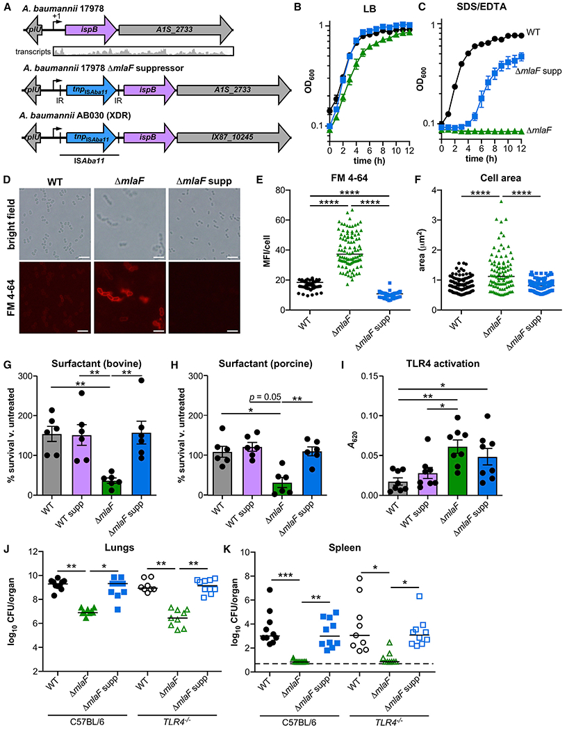
(A) Upstream of ispB and after its predicted transcription start site (+1), insertion ISAba11 includes inverted repeats (IR) and encodes a putative transposase.
(B and C) WT, ΔmlaF, and ΔmlaF suppressor (ΔmlaF supp) strains were grown in LB without (B) and with (C) 0.01% SDS and 0.175 mM EDTA (n = 3, data are means ± SEM).
(D) Live WT and ΔmlaF bacteria were incubated with the lipophilic fluorescent dye FM4-64 and imaged on agar pads. Scale bar is 5 μm.
(E and F) The mean fluorescence intensity (MFI) per cell (E) and the area per cell (F) were measured (n = 100 with median; significance by Kruskal-Wallis with Dunn’s multiple comparisons).
(G and H) WT, ΔmlaF and suppressor strain survival in Infasurf bovine (G) pulmonary surfactant and Curosurf porcine (H) pulmonary surfactant after 90 min (n = 6 from two independent experiments; data are means ± SEM; significance by one-way ANOVA with Tukey’s multiple comparisons).
(I) Activation of HEK-Blue TLR4 reporter cell line at multiplicity of infection 10−2 (n = 8; data are mean ± SEM; significance by one-way ANOVA with Tukey’s multiple comparisons).
(J and K) Wild-type (WT), ΔmlaF, and ΔmlaF suppressor strains were infected intranasally into C57BL/6 and TLR4−/− mice. At 36 h after infection, lungs (J) and spleens (K) were harvested, and bacterial burdens were enumerated (n = 9–10 with median; significance by Kruskal-Wallis with Dunn’s multiple comparisons). *p < 0.05, **p < 0.01, ***p < 0.001, ****p < 0.0001. See also Figure S3.