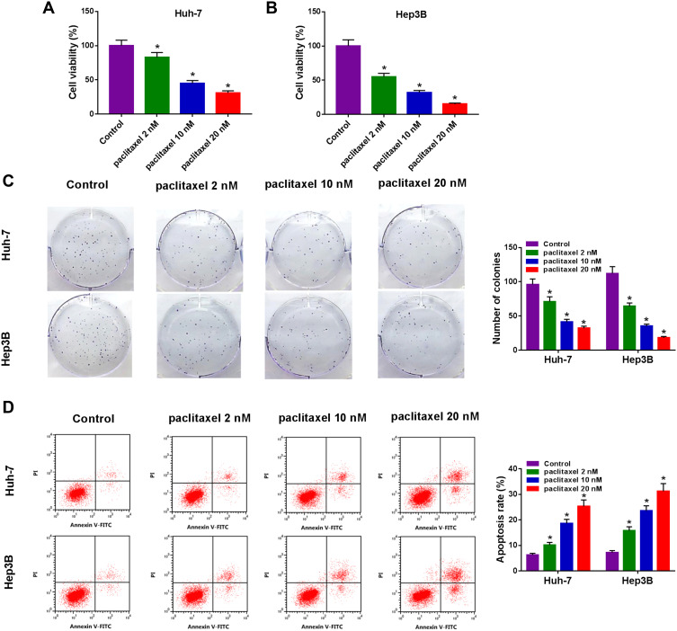
Figure 1.
Paclitaxel inhibited cell proliferation and contributed to apoptosis in HCC cells. Huh-7 and Hep3B cells were exposed to different concentrations of paclitaxel (2 nM, 10 nM and 20 nM). (A and B) CCK-8 assay was utilized to assess cell viability. (C) Colony formation assay was utilized to determine the number of colonies. (D) Cell apoptosis was examined with flow cytometry analysis. *P<0.05.

