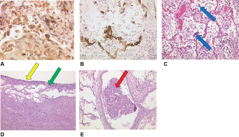
Fig. 5.

( A ) Lung tissue with IL-6 expression (brown colored stain) of plasma cells and alveolar macrophages (pneumocytes II, IL-6, Zytomed), ×80 magnification. ( B ) Lung tissue with MCP-1 expression (brown colored stain) in alveolar macrophages (pneumocytes II) and intra-alveolar fibrin (MCP-1, Santa Cruz), ×40 magnification. ( C ) The novel coronavirus disease (COVID-19) lung with activated intra-alveolar pneumocytes type II, some with virally altered multiple nuclei (blue arrow), H&E, ×20 magnification. ( D ) Left arteria carotis communis with endothelial damage (yellow arrow), necroses, fibrinous exsudation and inflammatory infiltrates of the intimal layer (green arrow), H&E, ×40 magnification. ( E ) COVID-19 lung. Early fibrin rich thrombus in a right sided pulmonary vein (red arrow) as a possible atypical origin of thromboembolic stroke, H&E, ×20 magnification. H&E, hematoxylin and eosin; IL, interleukin; MCP, monocyte chemoattractant protein.
