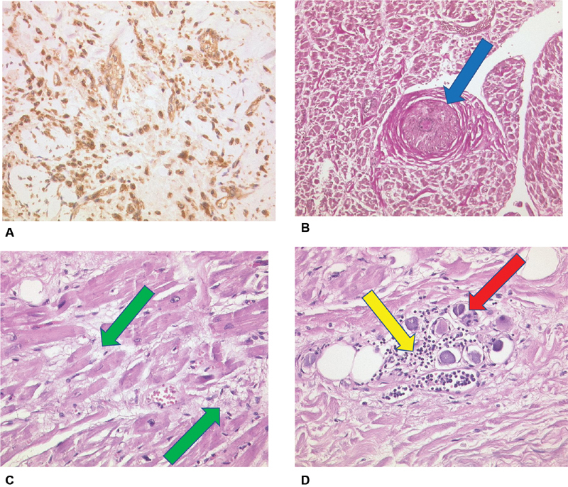
Fig. 6.

( A ) Thrombus in organization with IL-6-expression (brown colored stain) of endothelial cells, Fibroblasts and macrophages (IL-6, Zytomed, ×40 magnification). ( B ) Right atrium, stenosing microangiopathy “small vessel disease” (blue arrow), Elastika-van-Gieson (EvG), ×20 magnification. ( C ) Right atrium, fresh myocardial necroses (green arrows), H&E, ×40 magnification. ( D ) Right atrium, nerval ganglion cells with lymphocyte infiltration (yellow arrow) and viral alterations (red arrow), H&E, ×40 magnification. H&E, hematoxylin and eosin; IL, interleukin.
