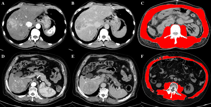
Fig. 1.
The Computed Tomography images of two different patients (fist: a, b, c; second: d, e, f) demonstrated two large hepatic lesions consistent with hepatocellular carcinoma due to the arterialization (arrows in a and d) coupled with wash-out of contrast media in the delayed phases (arrows in b and e). The diagnosis was confirmed by histology after surgical treatments in both patients. The evaluations at the level of the soma of the third lumbar vertebra by using dedicated free software revealed no sarcopenia in the first patient (c) and sarcopenia in the second one (f)

