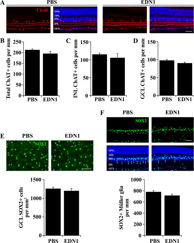
Fig. 5. EDN1 did not cause loss of amacrine cells or Müller glia.
a Retinal sections depicting choline acetyltransferase + (ChAT, red) amacrine cells and synaptic strata in the inner plexiform layer 28 days after PBS and EDN1 injection. Amacrine cell synaptic strata were morphologically indistinct 28 days after PBS and EDN1. b Quantification of total ChAT+ cells 28 days after PBS and EDN1. EDN1 injured retinas had similar numbers of ChAT+ amacrine cells compared to controls (ChAT+ cells/mm± SEM; PBS: 213.6 ± 4.0; EDN1: 196.4 ± 9.2, n ≥ 3 per condition, P = 0.117, two-tailed t test). ChAT+ amacrine cell numbers were similar in both the inner nuclear layer (INL; c INL ChAT+ cells/mm ± SEM; PBS: 115.3 ± 3.3; EDN1: 105.8 ± 11.3, n ≥ 3 per condition, P = 0.394, two-tailed t test) and the ganglion cell layer (GCL; d, GCL ChAT+ cells/mm± SEM; PBS: 98.3 ± 2.1; EDN1: 90.6 ± 2.2, n ≥ 3 per condition, P = 0.058, two-tailed t test). e Retinal flat mounts depicting SOX2+ amacrine cells in the ganglion cell layer 28 days after PBS and EDN1 injection. EDN1 did not cause significant loss of SOX2 + amacrine cells (SOX2 + cell survival ± SEM; PBS: 1263 ± 35.8; EDN1: 1204 ± 58.1; n = 7 per condition, P = 0.406, two-tailed t test). f Retinal sections depicting SOX2+ (green) Müller glia 28 days post-PBS and EDN1 injection. EDN1 did not appear to cause loss of SOX2+ Müller glia. Note: SOX2 labels a population of amacrine cells in the inner portion of the INL and in the GCL. Müller glia were distinguished from amacrine cells by location in the INL and by morphology in accordance with Surzenko et al.81. Arrowheads indicate SOX2+ amacrine cells in the INL that were not included in this quantification. (SOX2+ Müller glia/mm ± SEM; PBS: 783.1 ± 19.2; EDN1: 718 ± 22.0; n ≥ 3 per condition, P = 0.081, two-tailed t test). ONL outer nuclear layer; OPL outer plexiform layer; INL inner nuclear layer; IPL inner plexiform layer; GCL ganglion cell layer. Error bars, SEM. Scale bars, 50 μm.

