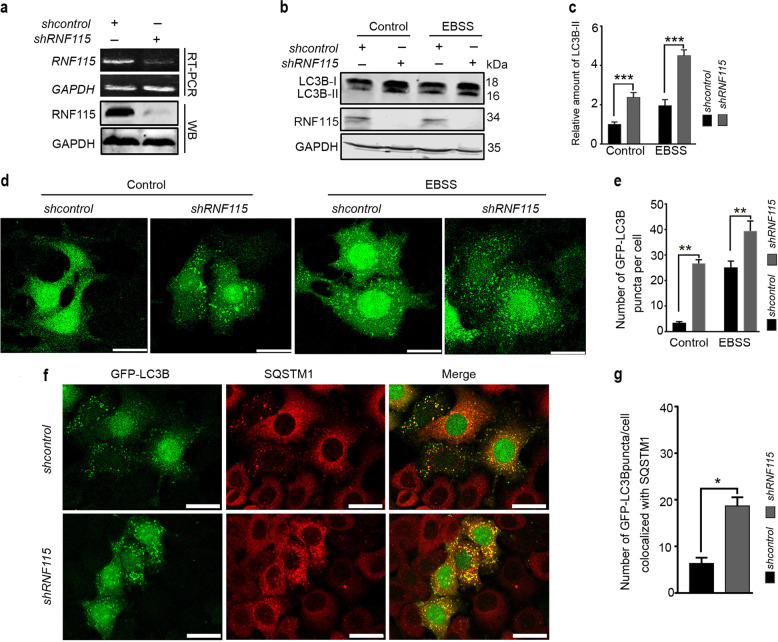
Fig. 1. Depletion of RNF115 casued autophagosome accumulation.
a RT-PCR and western blotting detected the levels of RNF115 in Hela cells. b, c HeLa cells were transfected with shcontrol or shRNF115 for 48 h, with or without EBSS for another 2 h, then LC3B-II levels were analyzed by western blotting. The relative amount of LC3B-II levels relative to GAPDH was analyzed. Average value in shcontrol-transfected cells without EBSS was normalized as 1. Data are means ± s.d. of results from at least three independent experiments. d, e Representative confocal microscopy images of GFP-LC3B distribution in stable GFP-LC3B Hela cells transfected with shcontrol or shRNF115 for 48 h, and treated with or without EBSS for another 2 h. The number of GFP-LC3B puncta/cell was calculated. Data are means ± s.d. of at least 50 cells scored. f, g Representative confocal microscopy images were shown in stable GFP-LC3B HeLa cells transfected with shcontrol or shRNF115 for 48 h, stained with anti-SQSTM1 antibody, and then observed by confocal microscopy. The number of GFP-LC3 puncta/cell colocalized with SQSTM1 aggregates was calculated. Data are means ± s.d. of at least 50 cells scored. Scale bar, 25 μm. *p < 0.05; **p < 0.01; ***p < 0.001.

