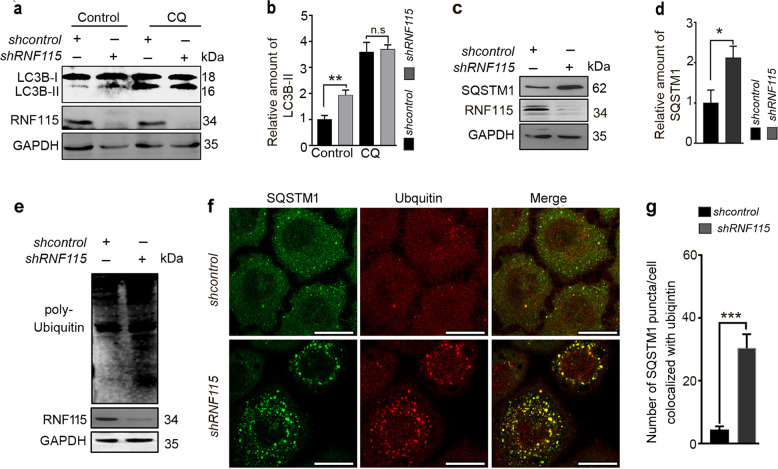
Fig. 2. Depletion of RNF115 blocks autophagic flux.
a, b HeLa cells were transfected with shcontrol or shRNF115 for 48 h, with or without CQ (50 μM) for 4 h, then the levels of LC3B-II were analyzed by western blotting. The quantification of LC3B-II levels relative to GAPDH was analyzed. Average value in shcontrol-transfected cells without CQ was normalized as 1. Data are means ± s.d. of results from at least three independent experiments. c, d HeLa cells were transfected with shcontrol or shRNF115 for 48 h, the SQSTM1 levels were analyzed by western blotting. The quantification of SQSTM1 levels relative to GAPDH was analyzed. Average value in shcontrol-transfected cells was normalized as 1. Data are means ± s.d. of results from at least three independent experiments. e Cells were treated as in c, the levels of ubiquitinated protein were detected by western blotting. f, g Hela cells were transfected with shcontrol or shRNF115 for 48 h, stained with anti-SQSTM1 and anti-ubiquitin antibodies, and observed by confocal microscope. The number of SQSTM1 puncta/cell colocalized with ubiquitin aggregates was calculated. Data are means ± s.d. of at least 50 cells scored. Scale bar, 25 μm. *p < 0.05; **p < 0.01; ***p < 0.001; n.s not significance.

