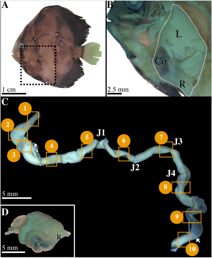
Figure 1. Characterization of gastrointestinal tract (GIT) sections of P. orbicularis and details of dissection.
(A) 56 dph P. orbicularis. Dashed square represents the limits of the dissection, see Fig. 1B. (B) View of the GIT inside P. orbicularis. Dashed white line delimits GIT. (C) Unrolled GIT. Yellow squares represent sections of interest for this work. These sections have been numbered from 1 to 10. J1 to J4: junction 1 to 4. The white arrowhead shows the valve between the colon and the rectum. (D) Isolated GIT after dissection. Co, colon; L, liver; R, rectum.

