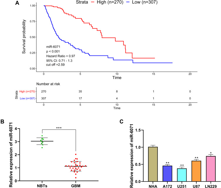
Figure 1.
miR-6071 expression was low in GBM and was associated with disease progression. (A) Kaplan–Meier analyses of the overall survival of The Cancer Genome Atlas (TCGA) cohort patients with high miR-6071 expression and those with low miR-6071 expression. (B) The expression of miR-6071 in GBM tissues was detected by qRT-PCR. (C) The expression of miR-6071 in GBM cells was detected by qRT-PCR. ***P < 0.001, vs Normal brain tissues (NBTs) group (B); *P < 0.05, **P < 0.01, vs NHA cells group (C).

