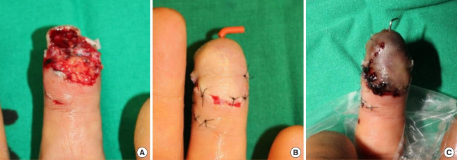
Fig. 3. Example of failure.
(A) Preoperative photo of a patient who was injured in a volar pattern. (B) Immediate postoperative photo; a pale color was observed, and the distal phalangeal bone fracture was fixed with K-wire. (C) Two weeks after the operation, total necrosis was observed. This case was judged as graft failure, as it required additional procedure(s) such as debridement.

