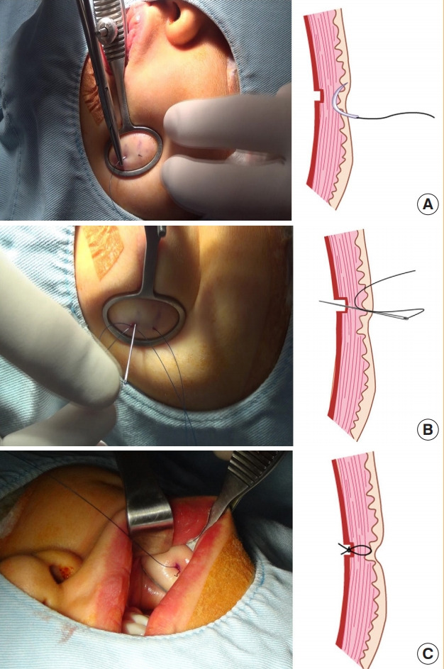
Fig. 1. Surgical method of dimple creation.

(A) From the site marked on the skin side (although some variation can exist depending on the desired dimple size), at average intervals of 0.3 to 0.8 cm, two sites were punctured vertically up to the dermis layer using 21-gauge needles. (B) Using a 4-0 nylon suture, from the punctured site in the skin side, a needle was passed through the dermis and inserted into the other puncture site. (C) Starting from the area where the needle emerged, the needle was passed through the incision site in the mucosa of the oral cavity. The two ends of the suture emerged from the incision site of the mucosa and were knotted.
