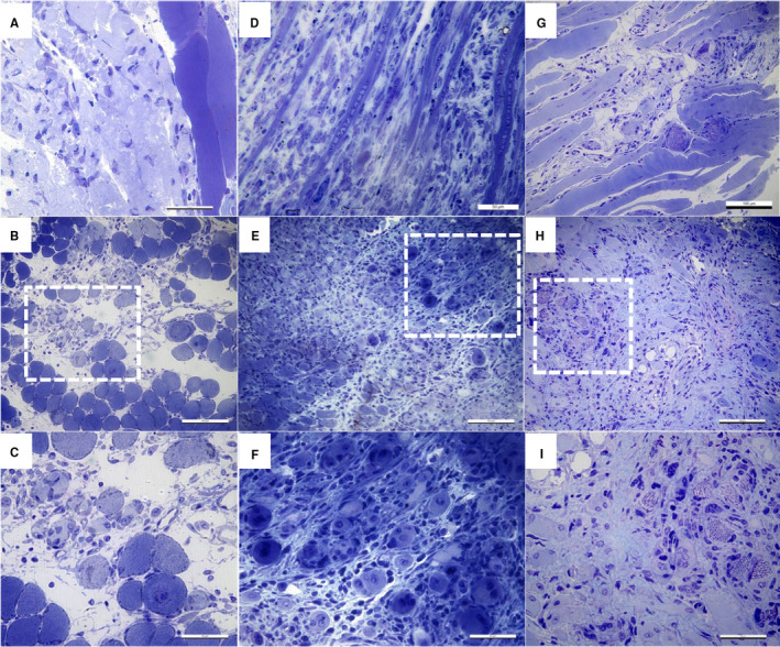
Figure 1.

Light microscopy on toluidine blue–stained semi‐thin sections of epoxy‐embedded injured gastrocnemius muscle. Representative images from three different mice. A‐C. 3 days post‐injury oedema and some necrotic fibres are observed along with a massive inflammatory infiltrate in the interstitial spaces around damaged fibres. D‐F. 5 days post‐injury, regenerating myofibres (myotubes with central nuclei) and inflammatory infiltrate are observed. In some areas, muscle necrosis is still present. G‐I. 14 days post‐injury, inflammatory infiltrate and collagen deposition were still detected at the injury site. A, D, G longitudinal sections; B, E, H cross sections; boxed areas are presented at a higher magnification in C, F and I, respectively
