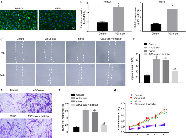
FIGURE 3.

miR‐486‐5p derived from ASC‐EVs accelerates HSFs proliferation and migration. A, ASC‐EVs internalized in HMECs and HSFs observed by fluorescence microscopy. EVs with green fluorescence were observed in recipient cells (×400). B, The expression of miR‐486‐3p in HMECs and HSFs treated with ASC‐EVs or PBS determined using RT‐qPCR after 3 h. C and D, The Migration rate of HSFs with different treatment measured and quantified using the scratch test. E and F, The Migration rate of HSFs with different treatment examined and quantified using Transwell assay (×400). G, Proliferation of HSFs assessed by CCK‐8 assay. *P < 0.05 compared with the control group. # P < 0.05 compared with the ASC‐EVs treatment. The measurement data were described as means ± standard deviation. Data between the two groups were analysed by unpaired t test whilst data amongst multiple groups were analysed by one‐way ANOVA followed by Tukey's test. Data at different time points were compared using repeated‐measures ANOVA with Bonferroni's test
