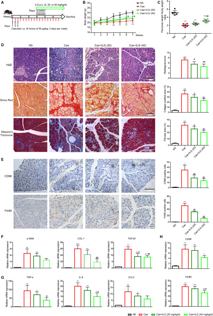
FIGURE 1.

Inhibitory actions of ILG against pancreatic fibrosis and inflammation. The flow diagram of caerulein‐induced experimental chronic pancreatitis (CP) and the intervention with ILG (A). The bodyweights of each group were monitored weekly over the experimental procedure (B). The relative pancreas weights were recorded at the end of the experiment (C). The histologic features of pancreases were exhibited by H&E, Sirius red and Masson's trichrome staining (D). Immunohistochemistry (IHC) staining for CD68 and F4/80 (brown, surface markers of macrophages) was performed on pancreatic paraffin sections (E). The histologic scores and quantitative analysis are presented, respectively, on the right side. Representative sections of pancreases were from at least five mice per group. qRT‐PCR analysis of the mRNA transcription of fibrogenic (F), pro‐inflammatory genes (G) and surface marker genes of macrophages (H). The data were expressed as relative fold changes over the values of the NS group and were all presented as mean ± SD of at least three independent experiments. *P < .05, **P < .01 compared with the NS group; #P < .05, ##P < .01 compared with the Cae group
