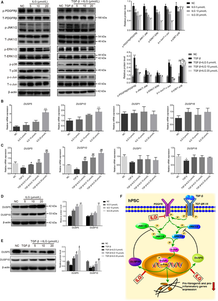
FIGURE 5.

ILG inhibits PSCs activation by regulating the MAPK signalling pathway. (A) WB analysis of the protein expression and activity levels of key factors from the MAPK signalling pathway (PDFGRβ, JNK1/2, ERK1/2, p38 and c‐Jun) in hPSCs with indicated ILG (left panel) or TGF‐β1 plus ILG (right panel) treatment for 4 h. The band signals were assessed using GeneTools software and statistically presented in the histogram on the right. (B and C) The mRNA expression level of DUSP5, DUSP10, DUSP1 and DUSP16 were analysed by qRT‐PCR in hPSCs treated with ILG alone (B) or TGF‐β1 plus ILG (C). The protein levels of DUSP5 and DUSP10 in hPSCs treated with ILG alone (D) or TGF‐β1 plus ILG (E) were analysed by WB and statistically presented on the right. All data were presented as mean ± SD of at least three independent experiments. *P < .05, **P < .01 compared with the control group; #P < .05, ##P < .01 compared with the TGF‐β1 group; ns: P > .05. (F) A schematic diagram illustrating the underlying mechanism by which ILG inhibits the expression of fibrogenic and pro‐inflammatory genes in hPSCs
