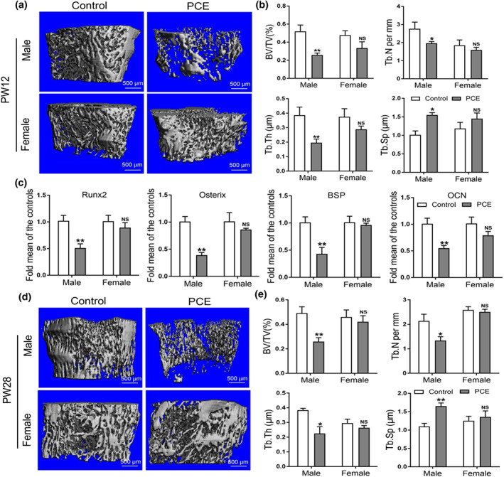
FIGURE 3.

Effects of prenatal caffeine exposure (PCE) on peak bone mass accumulation and osteogenic function in male and female adult offspring. (a) Representative micro‐CT images of femur from control and PCE offspring at 12 weeks old; (b) quantitative micro‐CT analysis of trabecular bone microarchitecture at 12 weeks old (n = 8 per group); (c) RT‐qPCR analysis of osteogenic marker genes expression, including Runx2, osterix, bone sialoprotein (BSP) and osteocalcin (OCN) in bone tissue at 12 weeks old (n = 8 per group). (d) Representative micro‐CT images of femur from control and PCE offspring at 28 weeks old; (e) quantitative micro‐CT analysis of trabecular bone microarchitecture at 28 weeks old (n = 8 per group); mean ± SEM, *P < 0.05 compared with the control; NS, no significance. Scale bar = 500 μm. BV/TV, bone volume/trabecula volume; Tb. N, trabecula number; Tb. Th, trabecula thickness; Tb. Sp, trabecula space
