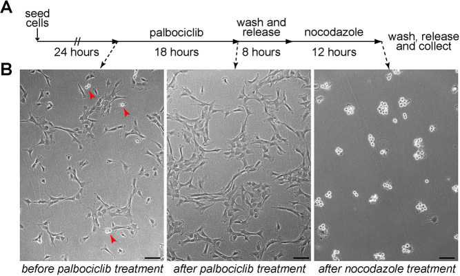
Fig. 1.
RPE-1 cells can be synchronized in mitosis using a palbociclib–nocodazole block. (A) Schematic diagram of the synchronization protocol. (B) Microscopy images of RPE-1 cells before and after palbociclib treatment (left and middle panels), and after nocodazole incubation (right panel). The red arrowheads in the left panel mark dividing cells. Note that the vast majority of cells are round dividing cells after nocodazole treatment. Scale bars: 100 µm.

