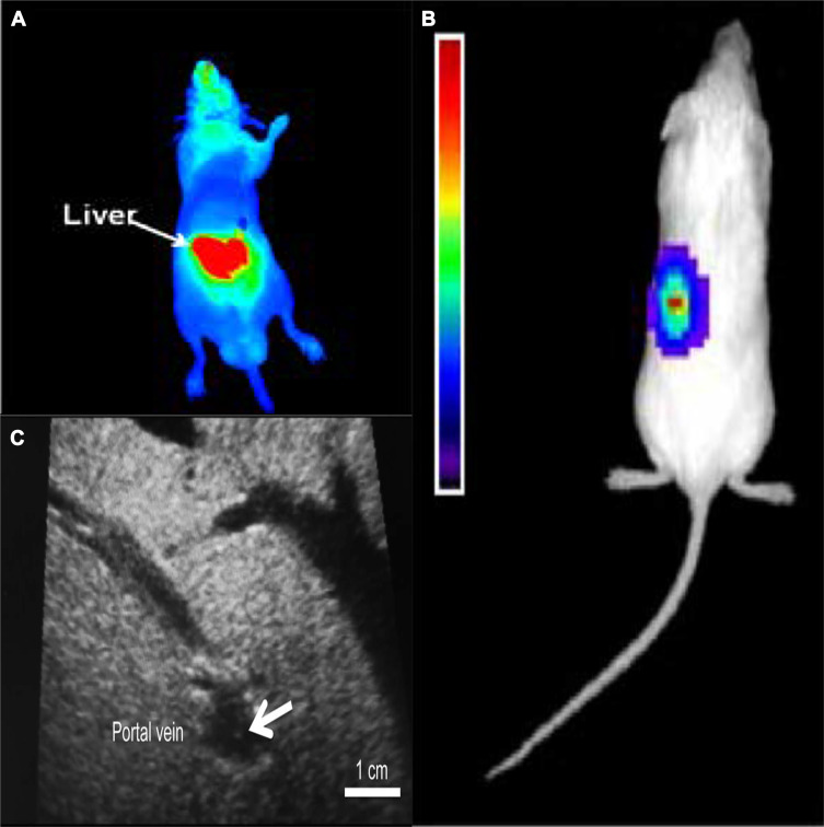
Figure 2.
Islets transplantation imaging of FI, BLI and US. (A) Transplanted rat islets were detected by PiF fluorescence imaging. Reprinted with permission from Kang NY, Lee JY, Lee SH, et al. Multimodal imaging probe development for pancreatic β cells: from fluorescence to PET. J Am Chem Soc. 2020;142(7):3430–3439. Copyright (2020) American Chemical Society.79 (B) 500 human islets were transduced with Adeno-CMV-Luc and implanted under the left kidney capsule of NOD-SCID mice. A representative CCD image 3 days postimplantation is shown. Reprinted from Mol Ther. 9(3). Lu Y, Dang H, Middleton B, et al. Bioluminescent monitoring of islet graft survival after transplantation. 428–435, Copyright 2004, with permission from Elsevier.65 (C) Intraoperative ultrasound findings of the portal vein. The transplanted islets appeared as hyperechoic clusters in the portal vein (arrows). Reproduced from Sakata N, Goto M, Gumpei Y, et al. Intraoperative ultrasound examination is useful for monitoring transplanted islets: a case report. Islets. 2012;4(5):339–342, reprinted by permission of the publisher (Taylor & Francis Ltd, hhtp://www.tandfonline.com).70

