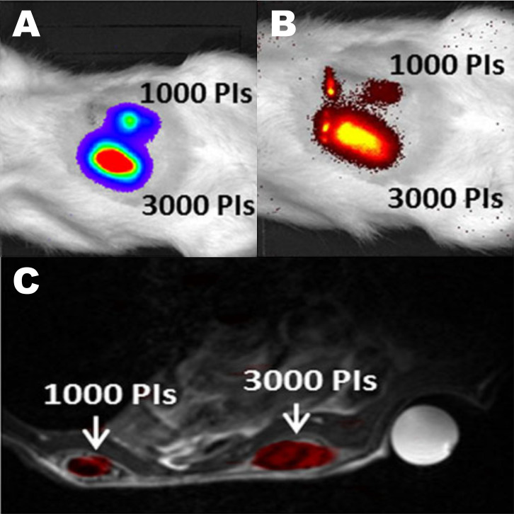
Figure 3.
Trimodal imaging of transplanted pancreatic islets in scaffolds. Representative (A) bioluminescence, (B) fluorescence, and (C) axial F-19/H-1 MR images of 3000 and 1000 pancreatic islets transplanted into scaffolds on days 4. Reproduced from Gálisová A, Herynek V, Swider E, et al. A trimodal imaging platform for tracking viable transplanted pancreatic islets in vivo: F-19 MR, fluorescence, and bioluminescence imaging. Mol Imaging Biol. 2019;21(3):454Y464. Creative Commons license and disclaimer available from: http://creativecommons.org/licenses/by/4.0/legalcode.11

