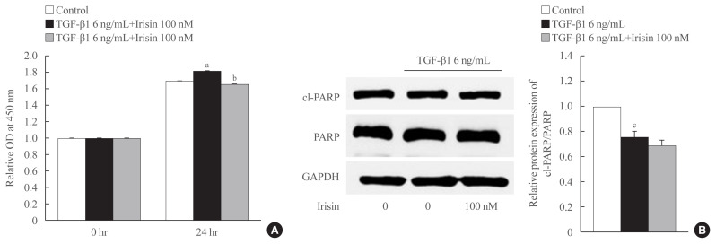
Fig. 3.
Irisin inhibits proliferation of activated hepatic stellate cell but is not involved in the apoptotic pathway. LX-2 cells were treated with transforming growth factor beta 1 (TGF-β1; 6 ng/mL) or TGF-β1 plus irisin (100 nM) for 24 hours. (A) Cell proliferation was measured using EZ-Cytox assay kit (DoGen). The optical density (OD) was measured at 450 nm. (B) Western blotting was used to analyze the apoptotic markers, and poly (ADP-ribose) polymerase (PARP) and cleaved-PARP (cl-PARP). Band intensities were measured using ImageJ software (National Institutes of Health) and are plotted to the right of the membrane images. Protein levels were normalized to glyceraldehyde 3-phosphate dehydrogenase (GAPDH). aP<0.01 vs. control; bP<0.01 vs. TGF-β1 only treatment (mean±standard error of the mean [SEM], n=6); cP<0.001 vs. control (mean±SEM, n=7).

