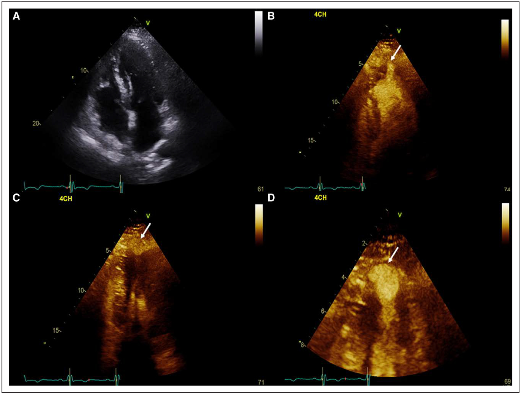
Figure 2. Echocardiography of apical hypertrophic cardiomyopathy with an aneurysm.

A, Apical 4-chamber view at end diastole with increased apical wall thickness. B, Apical 4-chamber view following administration of contrast in systole demonstrating a possible apical outpouching (arrow). C, Off-axis apical 4-chamber view of the apical outpouching at end systole (arrow). D, Zoomed in view of the apical aneurysm to clearly identify the wall (arrow; Movie I in the Data Supplement).
