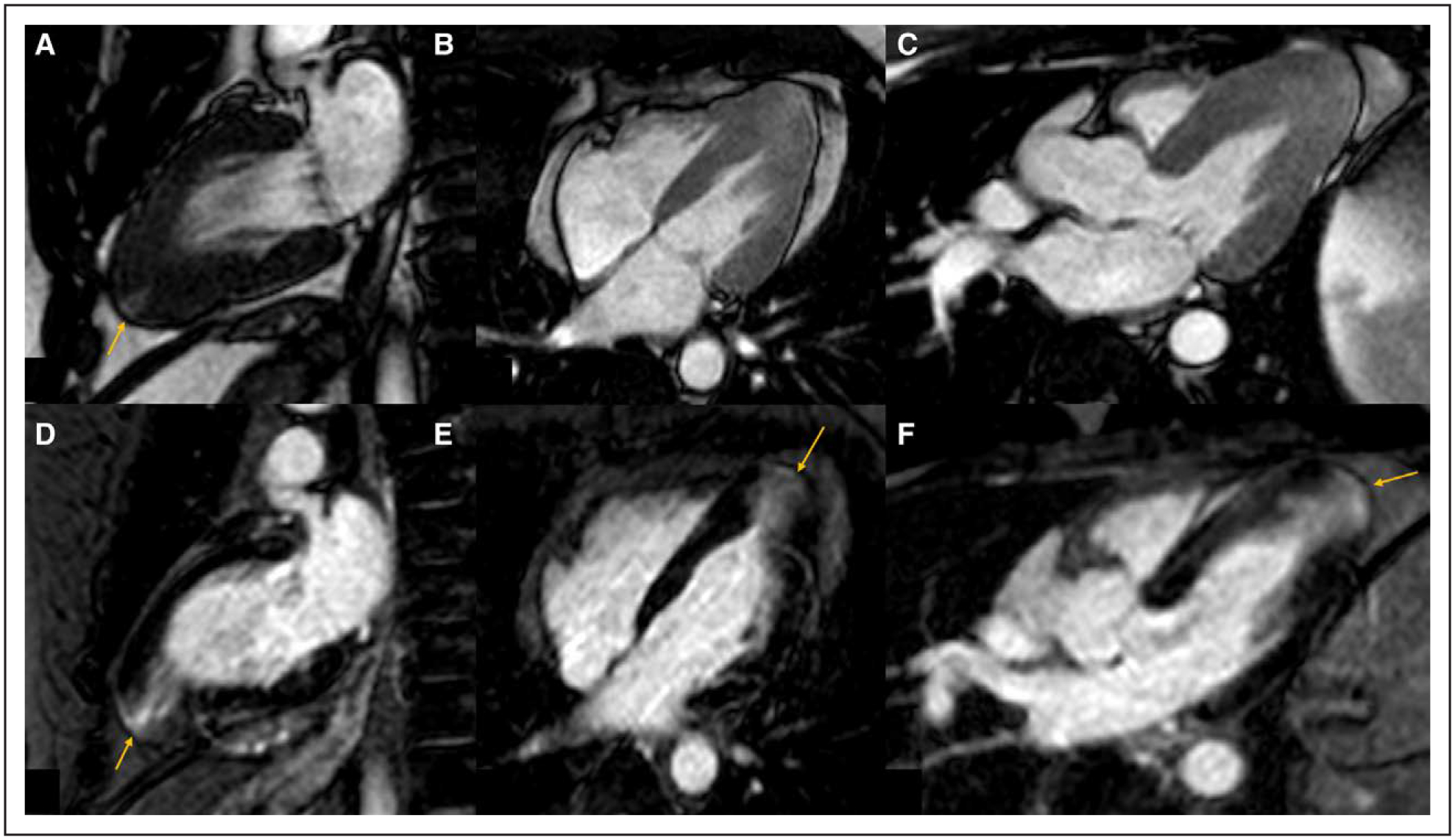
Figure 4. Cardiac Magnetic Resonance of apical hypertrophic cardiomyopathy with a small apical aneurysm.

A–C, Balanced steady-state free precession cine imaging in the vertical long axis, 4-chamber and left ventricular outflow tract (LVOT) view at end systole demonstrating increased apical wall thickness with a small apical aneurysm (arrow). D–F, Phase-sensitive inversion recovery imaging in the vertical long axis, 4-chamber and LVOT view demonstrating transmural late gadolinium enhancement of the apical aneurysm (arrow) and subendocardial to mid-myocardial late gadolinium enhancement of the apical segments.
