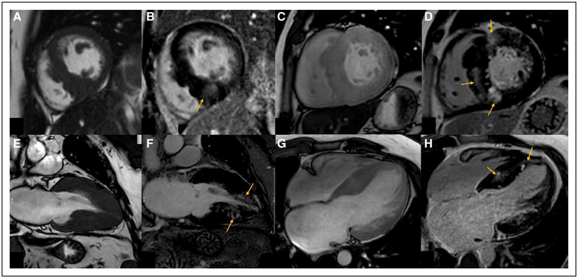
Figure 5. Variations in patterns of late gadolinium enhancement (LGE) and hypertrophy in patients with hypertrophic cardiomyopathy.

Patient 1: A, Balanced steady-state free precession (B-SSFP) cine imaging demonstrating asymmetrical septal hypertrophy predominantly affecting the mid inferoseptum. B, Phase-sensitive inversion recovery (PSIR) imaging in the short axis with mid-myocardial LGE in an area of maximal hypertrophy (arrow). Patient 2: C, B-SSFP cine imaging demonstrating asymmetrical septal hypertrophy. D, PSIR imaging with more extensive mid-myocardial LGE at the superior and inferior right ventricular insertion points and the mid septum (arrows). Patient 3: E, B-SSFP cine imaging in the vertical long-axis view demonstrating mid and apical hypertrophy. F, PSIR imaging with mid-myocardial LGE in the mid and apical segments (arrows). G, B-SSFP cine imaging in the 4-chamber view demonstrating mid to apical inferoseptal hypertrophy. H, PSIR imaging with mid-myocardial LGE in the mid to apical inferoseptum (arrows).
