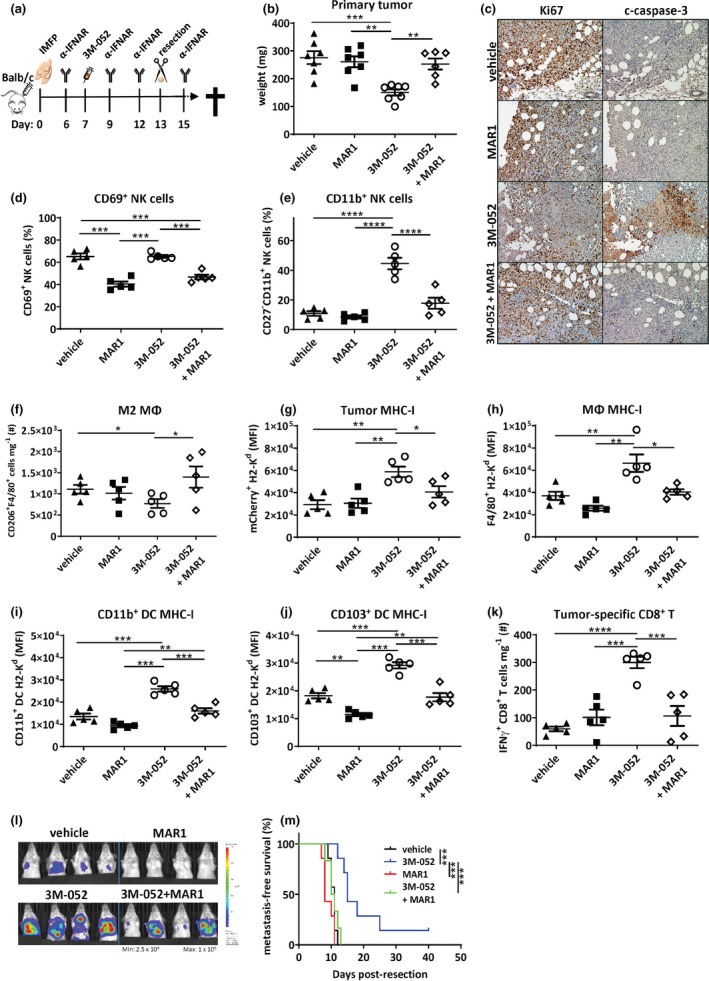
Figure 7.

Type I IFN signalling critical for 3M‐052 primary tumor and long‐term survival effects. (a) Experimental intervention timeline. BALB/c mice were injected with 1 × 10⁵ 4T1.2 cells into the 4th mammary fat pad, and palpable tumors were i.t. injected with vehicle or 3M‐052 on day 7 post‐inoculation. Mice were treated with four doses of isotype or MAR1‐5A3 mAb and primary tumors resected on day 13. Resected primary tumors were assessed for (b) weight (mg); (c) Ki67 and c‐caspase‐3 staining of formalin‐fixed primary tumors (20× magnification, scale bars = 100 µm, representative images shown); (d) proportion of CD69+TCRβ−NKp46+ cells; (e) proportion of CD11b+TCRβ−NKp46+ cells; (f) number of CD206+F4/80+ macrophages; and (g) tumor cell, (h) F4/80+ macrophage, (i) CD11b+CD11c+ and (j) CD103+CD11c+ DC MHC‐I (H2‐Kd) expression; and (k) number of IFN‐γ+TCRβ+CD8+ T cells. (l) Day 20 bioluminescence imaging of the thoracic cavity and (m) Kaplan–Meier survival curve comparing metastasis‐free survival. MΦ: macrophage. MFI: mean fluorescence intensity. Data are representative of one experiment. (b, l, m) n = 7 mice/group; (c) n = 2 mice/group; and (d–k) n = 5 mice/group. Statistical analysis was performed by one‐way ANOVA with post hoc Tukey's multiple comparison test and (m) the Mantel–Cox log‐rank test. Error bars are SEM. *P < 0.05; **P < 0.01; ***P < 0.001; and ****P < 0.0001.
