Fig. 3.
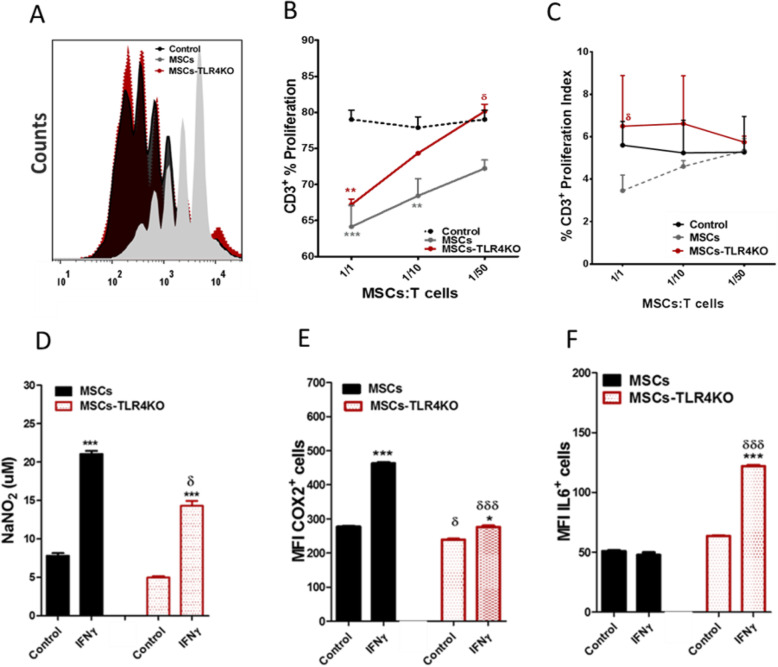
TLR4 expression mediates the immunosuppressive capacity of MSCs in vitro. CTV labeled splenocytes were cultured alone or with either MSCs WT or MSCs-TLR4KO at different MSCs: T cell ratio (1/1, 1/10, and 1/50) and activated with 1 μg/ml of ConA for 72 h. T cell proliferation was evaluated by FACS analysis. a Representative histograms for T cell proliferation with or without MSCs WT or MSCs-TLR4KO. Proliferation was calculated according to b frequency of total CD3+ T cells proliferation or c proliferation Index. d NO production was detected in the supernatants of MSCs WT or MSCs-TLR4KO using a modified Griess reagent. e COX2 and f IL6 expression was analyzed by flow cytometry in MSCs WT or MSCs-TLR4KO, pretreated or not with IFNγ for 48h. Data are expressed as mean ± SEM; n = 3, N = 3 biological replicates; *p < 0.05, ***p < 0.001 (inside each experimental group). δp < 0.05, δδδp < 0.001 (between each experimental group), derived by one-way ANOVA, Kruskal-Wallis ad-hoc post test

