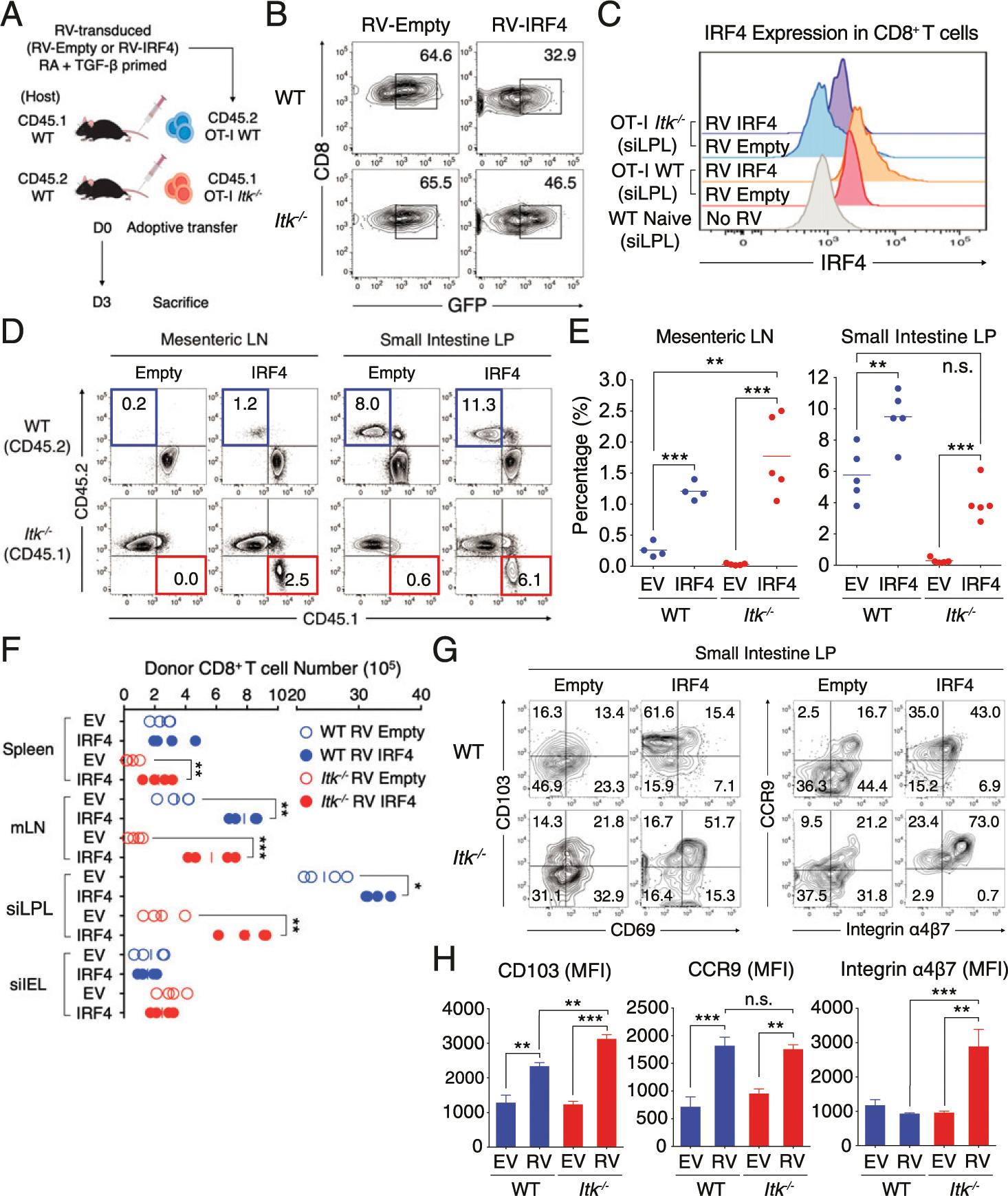
FIGURE 7. Enforced expression of IRF4 restores Itk−/− CD8+ T cell migration to the intestine.

(A) RV-transduced (Empty vector or IRF4) congenically labeled WT (CD45.2) and Itk−/− (CD45.1) CD8+ T cells were primed with RA and TGF-β for 48 h and then adoptively transferred (4.0 × 10⁷) to congenic WT naive host. (B and C) RV transduction efficiency (GFP+) (B) and IRF4 expression (C) of WT and Itk−/− CD8+ T cells from small intestine LP at day 3 of posttransfer are shown. No RV control shows the level of IRF4 expression in naive WT CD8+ T cells from siLPL. (D–F) Transferred T cells (WT, blue; Itk−/−, red) were isolated from mLN (left panel) and small intestinal LP (right panel) at day 3 posttransfer (D). Compilation data from four to five mice per each group from two to three independent experiments are shown at the right (E). Cell numbers of RV-transduced (Empty vector or IRF4) and RA- and TGF-β–primed donor OT-I cells in the spleen, mLN, siLPL and siIEL were enumerated in the congenic naive WT hosts at day 3 posttransfer (F). (G and H) The expression of CD69 versus CD103 (left) or integrin α4β7 versus CCR9 (right) in transferred T cells in small intestinal LP is shown (G). Mean fluorescence of each molecule was enumerated in the graph at the right (H). Statistical significance between individual groups was analyzed using unpaired or paired two-tailed Student t tests (*p < 0.05, **p < 0.01, ***p < 0.001).
