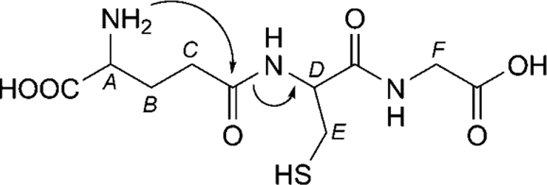
Fig. 1. Chemical representation of GSH.

Annotations denote chemical shift assignments in the NMR spectra: A: Glu-α, B: Glu-β, C: Glu-γ, D: Cys-α, E: Cys-β, F: Gly-α. Arrows denote the first step in the proposed degradation mechanism.

Annotations denote chemical shift assignments in the NMR spectra: A: Glu-α, B: Glu-β, C: Glu-γ, D: Cys-α, E: Cys-β, F: Gly-α. Arrows denote the first step in the proposed degradation mechanism.