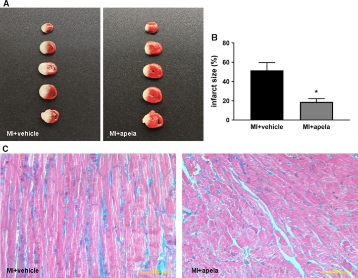
FIGURE 3.

Photographs showing representative TTC staining and histology of heart. The slices were incubated with 2,3,5‐triphenyltetrazolium‐chloride (TTC) for 30 min. Non‐infarcted myocardium was stained brick red, whereas infarcted tissue was unstained (A). The infarct size was measured and calculated as a percentage of the total area (B). Heart histology after Masson's trichrome staining of sections from each group (C).The data are presented as mean ± SD. *P < 0.05, vs. MI + vehicle group
