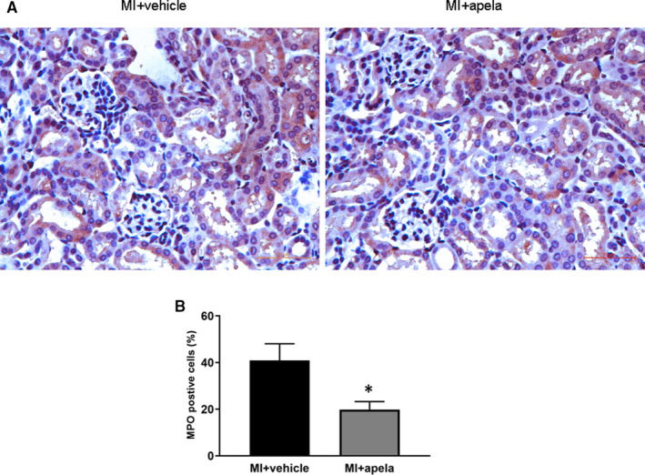
FIGURE 6.

Immunohistochemical (IHC) staining of MPO. Representative photomicrographs of MPO‐positive cells in kidney (A). Quantification of MPO‐positive cells in each group (B). The results were presented as mean ± SD. *P < 0.05, vs. MI + vehicle group
