
FIGURE 2.
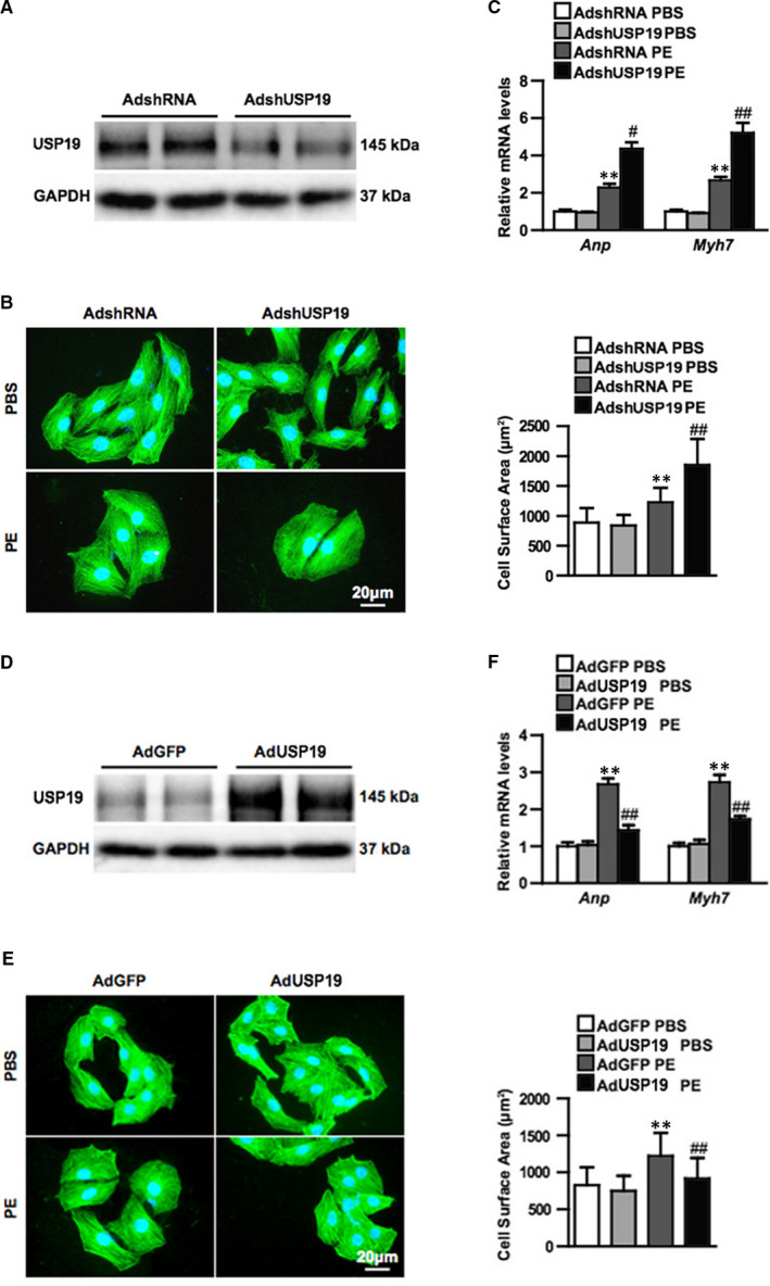
Ubiquitin‐specific protease 19 (USP19) attenuates phenylephrine (PE)‐induced cardiomyocyte hypertrophy in vitro. A, Western blot bands of USP19 levels in primary cultured cardiomyocytes infected with AdshRNA or short hairpin RNA (AdshUSP19) (n = 3 independent experiments). B, Representative microscopic images of cardiomyocytes infected with AdshRNA or AdshUSP19 and treated with phosphate buffered saline (PBS) or phenylephrine (PE). Cells were double stained of α‐actin (green) and 4‐6‐diamidino‐2‐phenylindole (blue). Scale bar, 20 μm. C, Quantitative results of the relative mRNA levels of atriopeptin (ANP) and myosin heavy chain 7 (Myh7, Above), and cross‐sectional area (Bottom) of cardiomyocytes infected with AdshRNA and AdshUSP19 in response to PBS or PE (n = 3 independent experiments, **P < 0.01 vs AdshRNA/PBS, ## P < 0.01 vs AdshRNA/PE). D, Western blot bands of USP19 levels in primary cultured cardiomyocytes infected with AdGFP or AdUSP19 (n = 3 independent experiments). E, Representative microscopic images of cardiomyocytes infected with AdGFP or AdUSP19 and treated with phosphate buffered saline (PBS) or phenylephrine (PE). F, Quantitative results of the relative mRNA levels of ANP and Myh7 (Above), and cross‐sectional area (Bottom) of cardiomyocytes infected with AdGFP and AdUSP19 in response to PBS or PE (n = 3 independent experiments, **P < 0.01 vs AdGFP/PBS, ## P < 0.01 vs AdGFP/PE), n.s. (no significance)