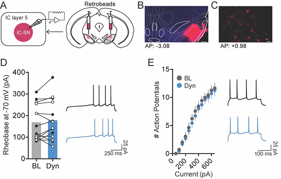
Figure 2. Activation of kappa opioid receptor (KOR) did not reduce the excitability of SN-projecting IC neurons.
(A) To label IC-SN neurons, fluorescent retrobeads were injected into the SN 7–10d before whole-cell recordings. (B) Representative images of retrobeads infused in the SN; scale bar, 250 μm and (C) retrobead labeling in IC-SN neurons; scale bar, 20 μm. Coordinates correspond to anterior-posterior (AP) position relative to bregma. (D) The minimum current required to elicit an action potential (rheobase) did not change from baseline (BL) following 10 min bath application of dynorphin A (Dyn, 300 nM); females data points are in open (white) circles, males are in closed (black) circles. (E) Dynorphin A did alter the number of action potentials fired per current injection step. Right panels in D and E show representative traces of current-injected firing in IC-SN neurons held at a common potential of −70 mV before (black) and after (blue) Dyn wash-on during a ramp protocol of 100 pA/1 s beginning at 200 pA (D) and at a 400 pA/250 ms current step (E). n = 3 mice/sex; BL, baseline; Dyn, dynorphin A

