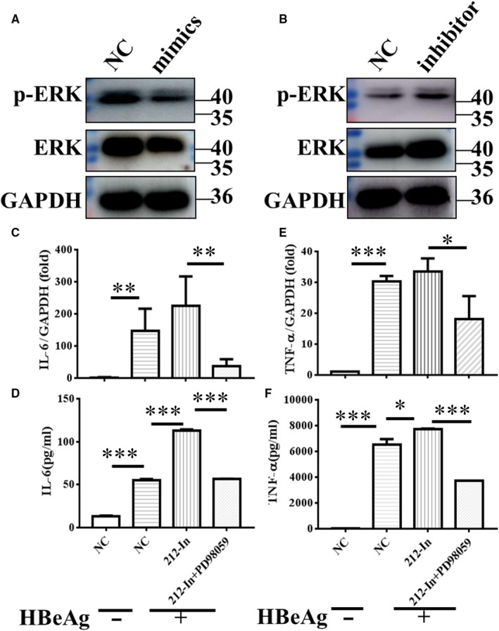
FIGURE 5.

miR‐212‐3p regulated HBeAg‐inducing inflammatory cytokines production via targeting MAPK1. Changes in non‐ phosphorylation and phosphorylation of ERK in RAW264.7 macrophages after transfection with miR‐212‐3p mimics or miR‐212‐3p inhibitor were determined with Western blot analysis (A, B). miR‐212‐3p inhibitor or negative control miRNA were transfected into RAW264.7 for 40 h, then cells were treated with DMSO or inhibitor of ERK (PD98059, 10 μmol/L) for 2 h, and cells were stimulated with HBeAg for 4 h. The expression and production of IL‐6 (C, D) and TNF‐α (E, F) were evaluated with q‐PCR and ELISA. Columns 1 and 2 represent the change of cytokine expression and secretion with or without HBeAg stimulation after transfection of NC, respectively (C‐F). Data are shown by three or four independent experiments (mean ± SD of triplicates in C‐F). *P < 0.05, **P < 0.01, ***P < 0.001
