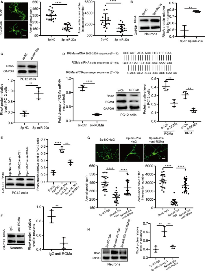
FIGURE 3.

Silencing miR‐20a‐5p inhibits primary hippocampal neuronal branching and axonal growth by regulating the RGMa‐RhoA pathway. A, Representative primary hippocampal neurons in the Sp‐miR‐20a and Sp‐NC groups captured by confocal laser scanning microscopy (bar = 25 µm). The axonal lengths of primary hippocampal neurons in Sp‐miR‐20a rats were significantly shortened compared with those in Sp‐NC rats (n = 20; two‐tailed t test). Neuronal morphology was assessed by Sholl analysis. Compared with that in the Sp‐NC group, the area under curve of the intersection numbers in the Sp‐miR‐20a group was significantly reduced (n = 20; two‐tailed t test), suggesting that silencing miR‐20a‐5p reduces neuronal branching of primary hippocampal neurons. B, miR‐20a sponge obviously decreased RhoA levels in primary hippocampal neurons (n = 3 independent cell culture preparations; two‐tailed t test). C, Down‐regulation of miR‐20a‐5p using a miRNA sponge distinctly increased RhoA expression in PC12 cells (n = 3 independent cell culture preparations; two‐tailed t test). D, RGMa‐siRNA targeting sites and sequences. RGMa mRNA and protein levels were down‐regulated by RGMa‐siRNA in PC12 cells (n = 3 independent cell culture preparations; two‐tailed t test). When the level of RGMa was decreased, the RhoA protein level was also decreased in PC12 cells (n = 3 independent cell culture preparations, two‐tailed t test). E, RGMa‐siRNA reversed the increase in RhoA induced by miR‐20a‐5p in PC12 cells (n = 3 independent cell culture preparations; one‐way ANOVA, Dunnett test). F, The functional RGMa antibody down‐regulated RhoA expression in primary hippocampal neurons (n = 3 independent cell culture preparations; two‐tailed t test). G, Representative primary hippocampal neurons in the Sp‐NC + IgG, Sp‐miR‐20a + IgG and Sp‐miR‐20a + anti‐RGMa groups captured by confocal laser scanning microscopy (bar = 25 µm). Compared with that in the Sp‐NC + IgG group, axonal growth in the Sp‐miR‐20a + IgG group was significantly shortened. When the functional antibodies were added to primary hippocampal neurons infected by miR‐20a sponge vectors, axonal growth was extended (n = 20; one‐way ANOVA, Dunnett test). Similar to the effects of the functional RGMa antibody on axonal growth, the functional RGMa antibody had the same influence on the area under the curve of the intersection numbers (n = 20; one‐way ANOVA, Dunnett test). H, The functional RGMa antibody reversed the up‐regulating effect of miR‐20a sponge on RhoA expression in primary hippocampal neurons (n = 3 independent cell culture preparations; one‐way ANOVA, Dunnett test). (**P < 0.01, ****P < 0.0001). All data represent the mean ± SD
