FIGURE 7.

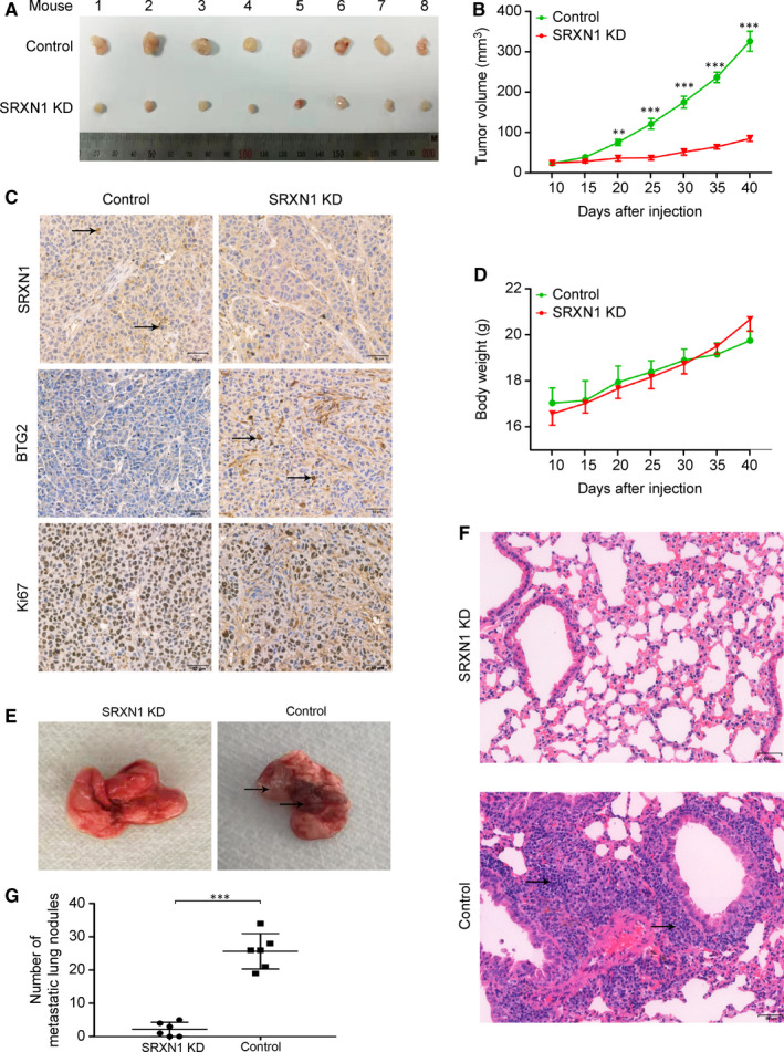
SRXN1 inhibits tumour growth and metastasis in vivo. A, Forty days after subcutaneous tumour cell inoculation, tumours were harvested and photographed (n = 8). B, The growth of tumours in the two groups was compared over time. Tumour volumes were calculated and recorded every 5 d. *** indicates P < 0.001; ** indicates P < 0.01. C, The bodyweight of each mouse was recorded every 5 d; the results are plotted against time. D, IHC analysis of tumour sections from the control and KD groups using antibodies against SRXN1, Ki67 and BTG2. The black arrow highlights the positive staining of related molecules in each panel. Scale bar: 50 μm. E, Lungs were harvested from mice in the control and KD groups 60 d after injection. Metastatic nodules were observed in the control group but not in the KD group. The black arrow indicates metastatic nodules in the lungs. F, HE staining of lung tissue samples from the control and KD groups. The black arrow indicates a metastatic nodule. Scale bar: 50 μm. G, Nodules in the lungs were counted and statistically analysed between control and KD groups. (n = 6)
