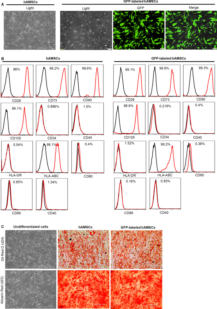
FIGURE 1.

Characterization of cell morphology and markers of hAMSCs and GFP‐labelled hAMSCs. A, Representative images of cultured hAMSCs and GFP‐labelled hAMSCs. B, Detection of surface markers in hAMSCs, GFP‐labelled hAMSCs (red) and in isotype controls (black) by flow cytometry. hAMSCs and GFP‐labelled hAMSCs were positive for CD29, CD90, CD73, CD105, HLA‐ABC, but negative for CD34, CD45, HLA‐DR, CD80, CD86 and CD40. C, Adipogenic differentiation of hAMSCs and GFP‐labelled hAMSCs was demonstrated by staining with oil red O, and osteogenic differentiation was demonstrated by Alizarin Red staining
