
FIGURE 2.
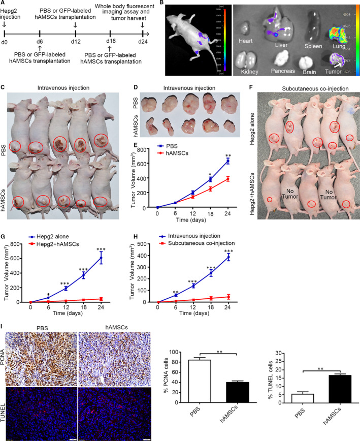
hAMSCs exhibit tumour‐homing and tumour‐suppressive properties in HCC mouse model. A, Experimental schematic of Hepg2‐induced hepatocellular carcinoma in the BALB/c nude mice model. The experiments were conducted in two groups including the PBS group and GFP‐labelled hAMSC group. B, Monitoring of hAMSCs tracking to tumour lesions in HCC mouse model on day 24 after Hepg2 transplantation by whole‐body fluorescent imaging assay. The results showed that GFP‐positive cells were detected around tumour site and in the tumour tissues. C and D, Gross observation of subcutaneous xenografts of Hepg2/PBS and Hepg2/hAMSCs intravenous‐injected nude mice. The red circles represent the tumour location. E, The average volume of hAMSC‐injected tumours was significantly smaller than that of PBS‐injected tumours at day 18 and day 24 (n = 5). F, Gross observation of subcutaneous xenografts of Hepg2 alone and Hepg2/hAMSCs co‐injected nude mice. The red circles represent the tumour location. G, The average volume of Hepg2/hAMSCs subcutaneous co‐injected tumours was significantly smaller than that of Hepg2 alone injected tumours at day 6, 12, 18 and 24 (n = 5). H, The average volume of Hepg2/hAMSCs subcutaneous co‐injected tumours was significantly smaller than that of hAMSCs intravenous‐injected tumours at days 6, 12, 18 and 24. I, Proliferation and apopsis of Hepg2 cells were tested by immunohistochemistry using antibodies against PCNA and TUNEL staining in PBS and hAMSCs intravenous‐injected tumour tissues. Results are shown as mean ± SD