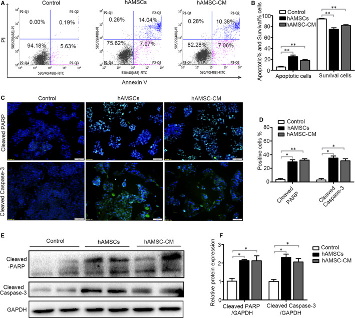
FIGURE 4.

Effect of hAMSCs and hAMSC‐CM on cell apoptosis in Hepg2 cells. A, Hepg2 cells were treated with normal medium (control), hAMSCs and hAMSC‐CM. The apoptosis of cells was assessed by FACS after 48 h of treatment. B, Quantitative analysis of the percentage of apoptotic cells and living cells as shown in A (n = 3). C, Effect of hAMSCs and hAMSC‐CM on cleaved‐PARP and cleaved caspase‐3 was detected in immunofluorescence assay. D, Quantitative analysis of the percentage of cleaved‐PARP positive and cleaved caspase‐3‐positive cells as shown in (C). E, The expression levels of cleaved‐PARP and cleaved caspase‐3 in Hepg2 cells of different groups were analysed by immunoblotting assay. F, Quantitative analysis of the expression of cleaved‐PARP and cleaved caspase‐3 in Hepg2 cells as in (E). Results are shown as mean ± SD
