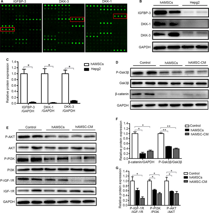
FIGURE 5.

hAMSC‐derived DKK‐3, DKK‐1 and IGFBP‐3 reduced activation of Wnt/β‐catenin and IGF‐1R/PI3K/AKT signalling pathway of Hepg2 cells. A, Representative array images are shown (n = 4). DKK‐3, DKK‐1 and IGFBP‐3 are highlighted with red boxes. B, Western blot analysis of DKK‐3, DKK‐1 and IGFBP‐3 protein levels in hAMSCs and Hepg2 cells. C, Quantitative analysis of the expression of IGFBP‐3, DKK‐1 and DKK‐3 in hAMSCs and Hepg2 cells as in (B). D, Hepg2 cells were treated with normal medium (control), hAMSCs and hAMSC‐CM. Western blot analysis of protein levels of β‐catenin, GSK3β and P‐GSK3β in Hepg2 cells of each treatment group. E, Western blot analysis of protein levels of IGF‐1R, P‐ IGF‐1R, PI3K, P‐ PI3K, AKT and P‐AKT in Hepg2 cells of control, hAMSCs and hAMSC‐CM group. F, Quantitative analysis of the expression of β‐catenin and P‐GSK3β in Hepg2 cells of different groups as in (D). G, Quantitative analysis of the expression of P‐ IGF‐1R, P‐PI3K and P‐AKT in Hepg2 cells of different groups as in (E)
