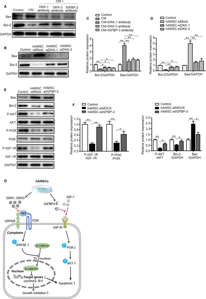
FIGURE 7.

DKK‐3, DKK‐1 and IGFBP‐3 derived from hAMSCs inhibit survival of Hepg2 cells through blocking Wnt/β‐catenin and IGF‐1R/PI3K/AKT pathway. A, Immunoblot analysis was performed on normal medium (Control), CM, CM + DKK‐1 antibody, CM + DKK‐3 antibody and CM + IGFBP‐3 antibody‐treated Hepg2 cell lysate using antibodies against GAPDH, Bax and Bcl‐2. B, Hepg2 cells were co‐cultured with normal medium (Control), hAMSC‐siMOCK, hAMSC‐siDKK‐1 and hAMSC‐siDKK‐3. Western blot analysis showed that the expression level of Bcl‐2 was increased and Bax was decreased in hAMSC‐siDKK‐3 group and hAMSC‐siDKK‐1 group when compared with hAMSC‐siMOCK group. C, Quantitative analysis of the expressions of Bcl‐2 and Bax in Hepg2 cells of different groups as in (A). D, Quantitative analysis of the expressions of Bcl‐2 and Bax in Hepg2 cells of different groups as in (B). E, Hepg2 cells were co‐cultured with normal medium (Control), hAMSC‐siMOCK and hAMSC‐siIGFBP‐3. The expression levels of Bax, Bcl‐2, P‐AKT, AKT, P‐PI3K, PI3K, P‐IGF‐1R, IGF‐1R and GAPDH in Hepg2 cells of different groups were determined by Western blot analysis. F, Quantitative analysis of the expressions of P‐IGF‐1R, P‐PI3K, P‐AKT, Bcl‐2 and Bax in Hepg2 cells of different groups as in (E). G, Schematic diagram of the extracellular and intracellular mechanisms of DKK‐3, DKK‐1 and IGFBP3 effect on the apoptosis and proliferation of Hepg2 cells. DKK‐3 and DKK‐1 secreted by hAMSCs can inhibit Wnt/β‐catenin signalling by sequestering LRP5/6, triggering apoptosis and inhibiting cell growth; IGFBP‐3 secreted by hAMSCs can inhibit IGF1 signalling by sequestering IGF1, resulting in cell apoptosis
