
FIGURE 5.
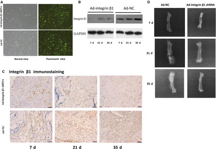
The infection efficacy of adenoviral carried integrin β1 shRNA (Ad‐integrin β1 shRNA) and adenoviral carried negative control (Ad‐NC) and representative X‐ray films of fractured femurs at 7, 21 and 35 d after fracture. A, To detect the infection efficacy of adenovirus in vitro, MCET3‐E1 cells were transfected and the green fluorescent view showed that the infected ratio was equal or greater than 95% compare to the normal view. B, The expression of integrin β1 in callus from the two groups was detected by Western blotting. The expression of integrin β1 in callus from Ad‐integrin β1 shRNA infected mice at 7, 21 and 35 d was obviously decreased compared with those in Ad‐NC groups (P < 0.05). C, Representative histological images of integrin β1 expression. The integrin β1 immunohistochemistry staining of callus in Ad‐integrin β1 groups at 7, 21 and 35 d were all significantly lower compared with those in Ad‐NC groups (P < 0.05). Scar bar = 50 μm. D, Representative X‐ray films of fractured femurs. 7 d, the two groups both showed disunion with clear fracture gaps and little periosteal callus. 21 d, the fracture lines were obscure in both the two groups, while the callus in Ad‐NC group seemed denser and bigger than that in Ad‐integrin β1 shRNA group. 35 d, the fracture line nearly disappeared and bony union was found in both the two groups