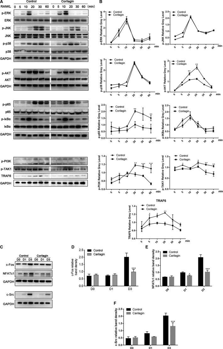
FIGURE 4.

Corilagin inhibited the activation of osteoclast formation via the NF‐κB and PI3K/AKT signalling pathways. A‐B, RAW264.7 cells were seeded into 6‐well plates and cultured with α‐MEM in the presence of 50 ng/mL M‐CSF overnight. The cells were pre‐treated with 2 μmol/L Corilagin or vehicle for 6 h and then stimulated with 100 ng/mL RANKL for 0, 5, 10, 20, 30 and 60 min, respectively. Cells were lysed, and lysates were detected using Western blotting with specific antibodies. C‐F, Corilagin impaired RANKL‐induced protein expression of NFATc1, c‐Fos and c‐Src in vitro. BMMs were treated with or without 2 μmol/L Corilagin in the presence of 50 ng/mL M‐CSF and 100 ng/mL RANKL for 0, 1 and 3 d, and total cell lysates were analysed with the same way above. All results are performed as mean ± SD; *P < .05, **P < .01, ***P < .001 vs the control group
