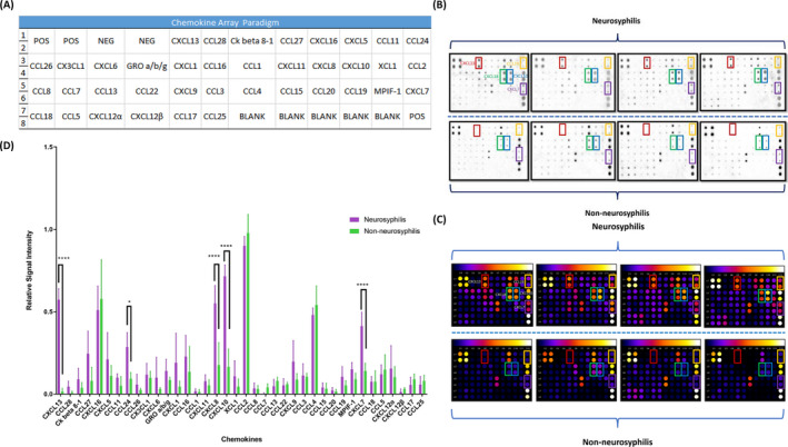
FIGURE 1.

Chemokine array performed on four patients with neurosyphilis and four with non‐neurosyphilis. A, Full chemokine array paradigm. B, Digital images taken with a chemiluminescence imaging system. C, Signal intensities recognized by ImageJ with Protein Array Analyzer plugin. D, Relative signal intensity of 38 chemokines in four patients with neurosyphilis and four with non‐neurosyphilis. CXCL13 is indicated by the red rectangle, CCL24 by the yellow rectangle, CXCL8 by the green rectangle, CXCL10 by the blue rectangle, and CXCL7 by the purple rectangle
