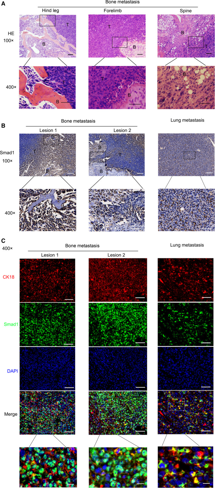
Figure 2.

BMP activation was higher in bone metastasis than in lung metastasis of Lewis lung carcinoma. A, Lewis lung carcinoma cells (LLCs) were injected into tail veins of C57/BL6 mice, resulting in lung metastasis and bone metastasis. Representative HE staining of metastatic bone tumours from limbs or spines was shown. Scale bars, 100 μM. Regions in the rectangles were magnified to 400×. Scale bars of the 400 × photos were 50 μM. T: Tumour; B: Bone. B, Representative Smad1 immunohistochemical staining of metastatic bone tumours and metastatic lung tumours metastasis tissues. Scale bars of the 100 × photographs were 100μM. Regions in the rectangles were magnified to 400×. Scale bars of the 400 × photographs were 50μM. T: Tumour; B: Bone. C, Representative immunofluorescence staining of Smad1, CK18 and DAPI. Scale bars of the 400 × photos were 50 μM. Regions in the rectangles were magnified, scale bars: 10 μM. Red: CK18‐TRITC; Green: Smad1‐FITC; Blue: DAPI
