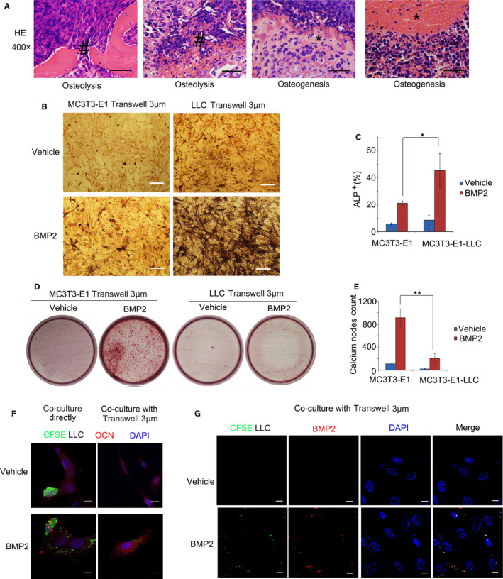
Figure 6.

BMP2 regulated the osteolytic and osteoblastic mechanisms of bone metastasis in Lewis lung carcinoma. A, 1 × 10⁶ LLCs were injected into tail veins of C57BL/6 mice, resulting in bone metastasis. Representative HE staining of bone metastasis tissues from limbs or spines was shown. Lysed bones (#). Immature bones (*). Scale bars, 50 μM. B, LLCs or MC3T3‐E1 cells were placed on the upper layer of cell culture insert with polycarbonate membrane (Transwell@, 3.0μm pore size, Corning). MC3T3‐E1 cells were cultured below in culture media with or without 200ng/mL BMP2 for seven days. ALP staining for MC3T3‐E1 cells cultured below was conducted. Representative photographs were shown. Scale bars, 10μM. C, Average ALP+ cell numbers of at least three fields in (C) were shown. The P value was based on Student's t test. (*P < .05, **P < .01). D, LLCs or MC3T3‐E1 cells were treated the same way as shown in (B) for 14 days. MC3T3‐E1 cells cultured below were stained with 1% Alizarin red (pH = 4.2). Representative photos were shown. E, Average calcium node numbers in (F) of at least three wells were shown. The P value was based on Student's t test. (*P < .05, **P < .01). F, 3 × 10⁴ MC3T3‐E1 cells were co‐cultured with 3 × 10⁴ CFSE‐labelled (green) LLCs directly (left). 3 × 10⁴ MC3T3‐E1 cells were seeded into the wells of the 6‐well co‐culture plates (Corning), and 3 × 10⁴ CFSE‐labelled (green) LLCs were seeded on the upper layer of Corning cell culture insert with polycarbonate membrane (Transwell@, 3.0 μm pore size, Corning) with 200 ng/mL BMP2 or vehicle (right). OCN‐TRITC (red) was stained to show MC3T3‐E1 cells. DAPI (blue) was stained to show the nucleus. Representative immunofluorescence photographs were shown. Scale bars, 10 μM. G, 3 × 10⁴ MC3T3‐E1 cells and 3 × 10⁴ CFSE‐labelled (green) LLCs were seeded on the Corning transwell the same way as shown in (F). BMP2 were stained (Red). DAPI (blue) was stained to show the nucleus. Representative immunofluorescence photographs were shown. Scale bars, 10 μM
