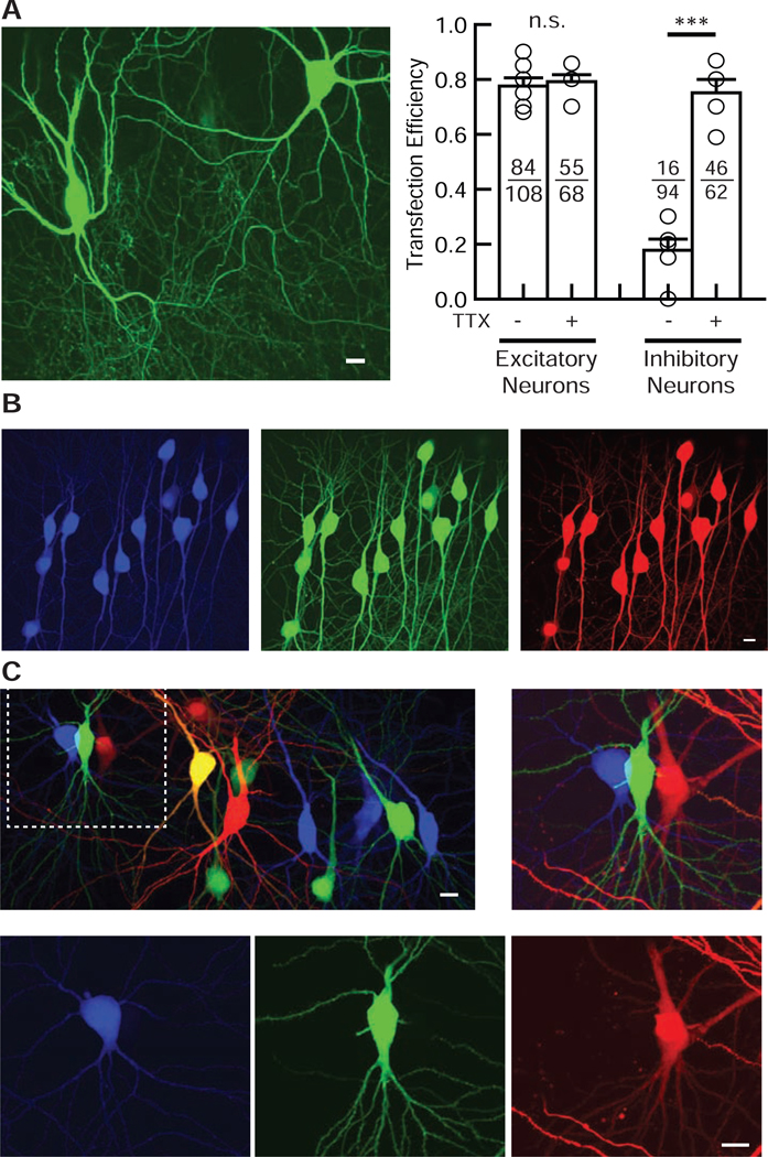
Figure 2. Validation of the electroporation method in both excitatory and inhibitory neurons.
(A) TTX improves the transfection efficiency in inhibitory but not excitatory hippocampal neurons. (Left) Confocal image of EGFP-transfected interneurons in the hippocampal CA1 oriens region. Interneurons were identified by morphology and anatomy. (Right) Summary bar graph of the transfection efficiency of excitatory and inhibitory neurons with or without TTX. Each symbol represents the transfection efficiency obtained from one organotypic slice culture. Numbers in each bar represent the numbers of transfected (numerator) and electroporated (denominator) neurons. Student’s t-test: n = 5 – 7 slice cultures from 2 mice for each condition. Data shown are means ± SEM. ***p<0.001, n.s.: not significant. (B) Confocal images of Tag-BFP, EGFP, and DsRed2 triple gene transfection by electroporation in hippocampal CA1 pyramidal neurons. Internal solution contained 33 ng / μl of each plasmid. Note that all transfected pyramidal neurons expressed all three co-transfected genes. (C) Confocal images of three non-overlapping gene transfections using Tag-BFP, EGFP and DsRed2 electroporated in hippocampal CA1 pyramidal neurons. Three different gene plasmids, Tag-BFP, EGFP, and DsRed2, were loaded into glass pipettes one at a time, and three sets of electroporations were done sequentially in different neurons. The middle left neuron that showed up as yellow was electroporated with both EGFP and DsRed2, leading to their co-expression.

