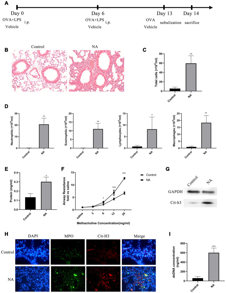
Figure 1.
NETs were generated in a mouse model of neutrophilic asthma (NA). (A) Flow chart for generation of NA mice. (B) HE staining of lungs showed prominent inflammatory cell infiltration around the bronchi and blood vessels of NA mice (200X). (C–E) Total cells, differential cell count, and total protein in the BALF supernatant of NA mice were measured. (F) Airway resistance was measured in NA mice after treatment with different concentrations of methacholine. (G) Cit-H3 expression was measured by western blotting. (H) Cit-H3 and MPO expression was measured by immunofluorescence (400X). (I) dsDNA concentration in the BALF supernatant of NA mice was detected by PicoGreen analysis. *: P<0.05, **: P<0.01, ***: P<0.001 vs control group.

