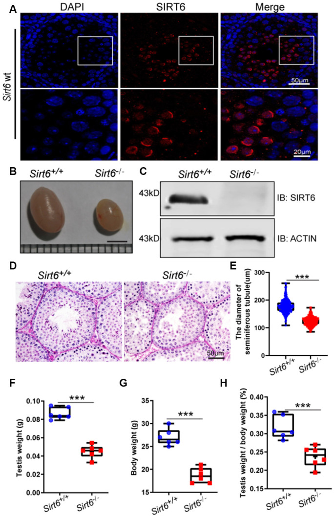
Figure 1.

SIRT6 protein expression and localization in mice testes. (A) Testicular sections of Sirt6+/+ stained for SIRT6 (red) and DAPI (blue). SIRT6 is localized in the spermatids. 8-week mice, n=3. (B) The testes of Sirt6-/- mice were smaller than those of the Sirt6+/+ mice. 8-week mice, n=6, scale bar=3 mm. (C) SIRT6 protein levels were dramatically reduced in the testes of the Sirt6-/- mice. ACTIN was used as the loading control. 8-week mice, n=3. (D) Histological analysis of Sirt6+/+ and Sirt6-/- mice seminiferous tubules by PAS-hematoxylin staining. 8-week mice, n=4. (E) The diameter of the seminiferous tubules in Sirt6-/- mice was smaller than that in control mice. Sirt6+/+, 177.43±2.36μm; Sirt6-/-, 123.94±1.92μm. 8-week mice, n=6; 300 seminiferous tubules were used for each group. Data are presented as mean ± SEM. ***P < 0.001. (F) Quantification of testis weight of the Sirt6+/+ and Sirt6-/- mice. The testis weight of Sirt6-/- mice was significantly reduced. Sirt6+/+, 8.60±1.30% g; Sirt6-/-, 4.49±1.40% g. 8-week mice, n=6. Data are presented as mean ± SEM. ***P < 0.001. (G) Quantification of body weight of the Sirt6+/+ and Sirt6-/- mice. The body weight of Sirt6-/- mice was reduced. Sirt6+/+, 27.00±0.55 g; Sirt6-/-, 18.67±0.30g. 8-week mice, n=6. Data are presented as mean ± SEM. ***P < 0.001. (H) The testis weight/ body weight of Sirt6-/- mice was also reduced. Sirt6+/+, 0.32±0.07%; Sirt6-/-, 0.04±0.07%. 8-week mice, n=6. Data are presented as mean ± SEM. ***P < 0.001.
