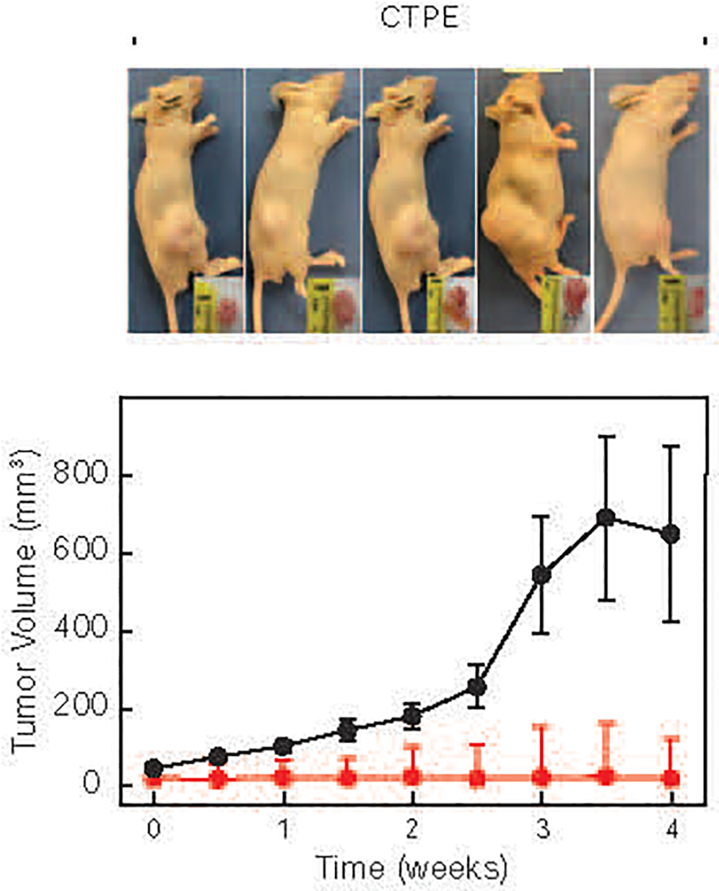
Figure 2. Formation of tumors in xenografts in nude mice.

RWPE-1 (red line) and CTPE (black line) cells were injected into nude mice and tumor growth monitored twice a week. Tumor and mouse images were obtained 31 days after injection.

RWPE-1 (red line) and CTPE (black line) cells were injected into nude mice and tumor growth monitored twice a week. Tumor and mouse images were obtained 31 days after injection.