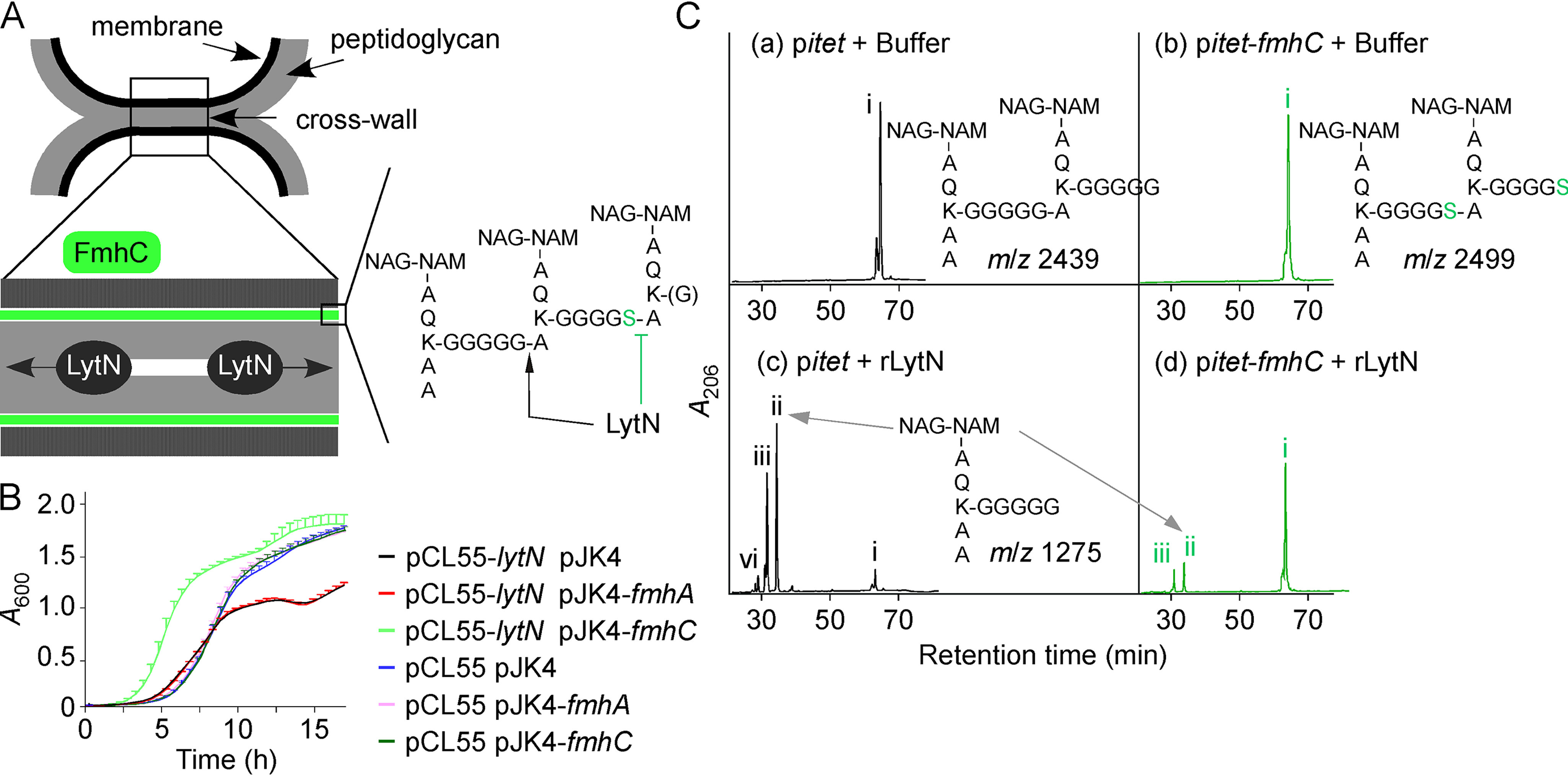
Figure 4.

FmhC acts as an immunity factor for LytN. A, schematic of dividing daughter cells illustrating the location of the cell wall envelope, membrane (black), and peptidoglycan (gray). The cross-wall structure has been expanded to show LytN cleaving peptidoglycan. The model postulates that FmhC synthesizes peptidoglycan (shaded in green) that is resistant to LytN hydrolysis. The proposed structure of this cross-linked peptidoglycan is shown with sites cleaved by LytN (black arrow) or refractory to LytN cleavage (green inhibition sign). Green and blue hexagons: GlcNAc and N-acetylmuramic acid, respectively. B, the growth rate of S. aureus Newman pCL55 and Newman pCL55-lytN carrying pJK4, pJK4-fmhA, or pJK4-fmhC was interrogated by monitoring changes in A600 over 16 h at 37 °C. Cultures were grown in the presence of anhydrotetracycline and IPTG inducers. C, purified dimeric peptidoglycan fragments obtained from Newman carrying control vector pitet or pitet-fmhC, as shown in Fig. 2B, were treated with buffer (insets a and b) or rLytN (insets c and d). Reaction products were separated by HPLC. Peaks denoted with Roman numerals (i–iv) were subjected to MALDI-TOF analysis and the corresponding m/z values are listed in Table 2. Predicted structures of peptidoglycan fragments i (panels a and b) and ii (panels c–d) are shown on the figure.
